DNA Origami Nanosyringes
A syringe stitched from DNA strands
Imagine needing to slip a carefully chosen message into a sealed bubble without tearing the bubble apart—and then closing the door behind you. Nature already does something like this. Certain bacteria fire contractile injection systems, protein machines that punch target membranes and deliver cargo in one decisive motion.
Engineering that same mix of grip, push, and precision at the nanoscale has been stubbornly hard. Now a team led by Na Liu at the University of Stuttgart and the Max Planck Institute for Solid State Research, working with collaborators including groups in Hangzhou, has built a fully synthetic answer from DNA origami: a programmable DNA origami nanosyringe, or DOS.
It is not a miracle machine. It is something quieter and more delightful—a modular, reversible device that binds a lipid membrane, drives a needle downward in controlled steps when fed short DNA “fuel” strands, and can pull the needle back so the membrane reseals. Along the way it can ferry tethered cargo and switch on biochemical reactions inside cell-sized compartments.
Two beams, one gold hinge, a sliding needle
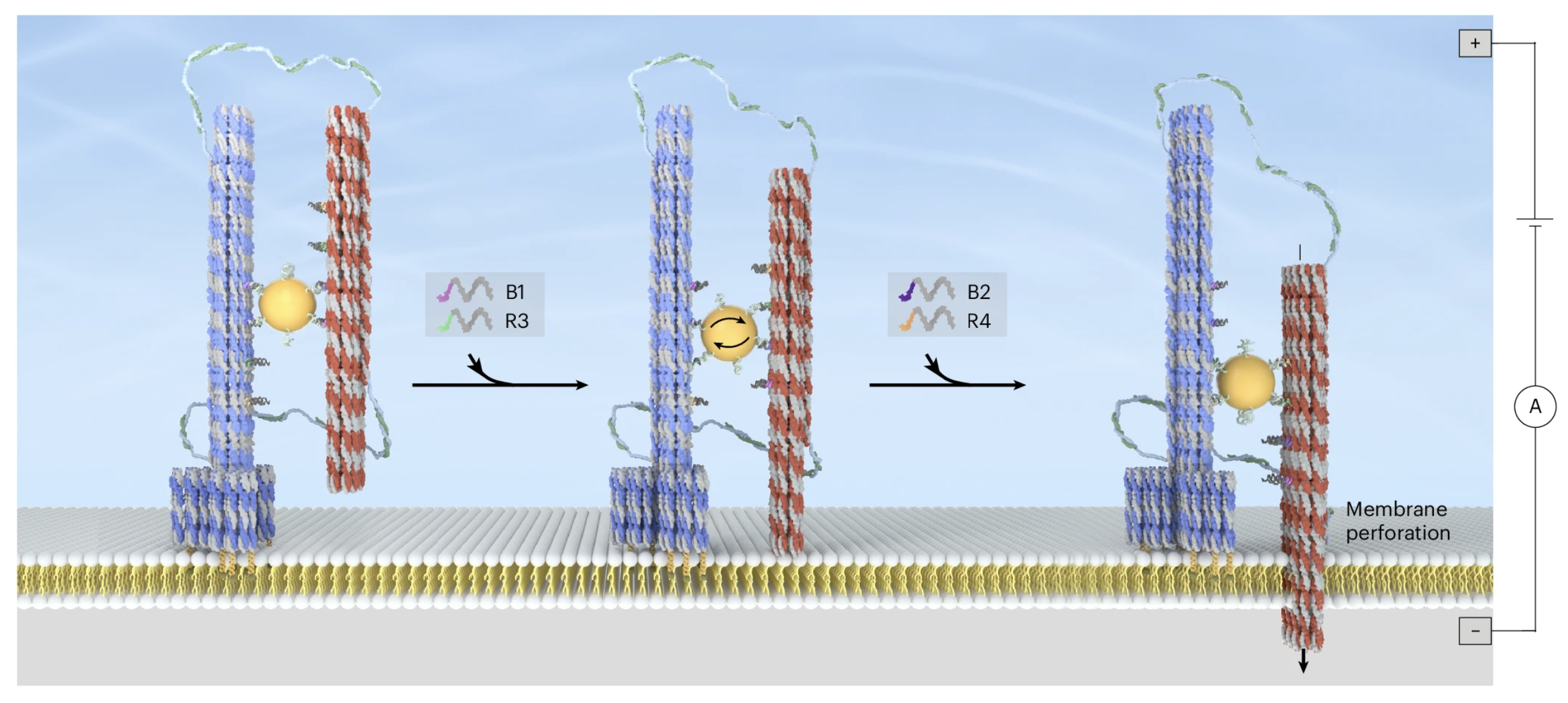
The core looks almost architectural. Two DNA origami bundles—each a roughly 70-nanometer bundle of DNA helices—sit antiparallel and are crosslinked by a 10-nanometer gold nanoparticle. A continuous scaffold strand helps keep them aligned while still allowing motion. Four rows of footholds line the inner faces of the bundles. The gold particle grips two rows on each side; DNA fuel strands then perform toehold-mediated strand displacement—a molecular swap in which one DNA strand peels another off a binding site and hands the particle a new foothold. Each successful step shifts the bundles by about 14 nanometers; two steps give a maximum relative slide of 28 nanometers.
One bundle carries a multihelix base studded with twelve cholesterol-tagged DNA anchors. Cholesterol loves lipid membranes the way oil loves oil, so the base settles onto supported bilayers or vesicle surfaces without clumping. The other bundle is the needle. Its honeycomb lattice of helices encloses three slender central channels, each about two nanometers across. Seven sites at the tip wait for cargo.
Once the base is parked, fuel strands send the needle downward. Transmission electron microscopy captures the shift from a symmetric resting pose to an asymmetric, membrane-engaged one. Electrophysiology on free-standing bilayers tells the functional story: binding alone produces no current leak; after fuel is added, ionic current rises in steps as a conductive pore opens. The pore stays open for more than an hour under continuous recording, and the current–voltage curve stays linear—consistent with a clean path through those central channels. Onset timing even sits in the same ballpark as familiar natural pores such as OmpF and aerolysin.
Piercing synthetic cells—and stitching them shut again
On giant unilamellar vesicles—soft, cell-sized lipid bubbles—the nanosyringes light up under the confocal microscope when labeled with fluorescent dyes. A small dye, sulforhodamine B, barely enters before actuation. After the needle slides, it pours in. Swap the dye for a bulkier or more negatively charged cousin, or for a three-kilodalton dextran roughly three nanometers across, and transport drops sharply. That size- and charge-selectivity is a quiet reassurance: most traffic is going through the designed channels, not through ragged tears.
Harder membranes are harder to pierce. When the team varied lipid recipes to raise mechanical resistance, penetration efficiency fell—honest evidence that the needle is doing real mechanical work against a real barrier.
The reverse stroke may be the most elegant part. Add fuel for upward sliding and bilayer current returns toward baseline. Fluorescence recovery after photobleaching—a way of watching how freely labeled devices wander in the membrane—slows when the needle is spanning the bilayer, then speeds up again after retraction, as if the device has unlatched. Dye influx also drops after the pull-back. A control needle whose channels were deliberately packed shut barely lets dye through at all, underscoring that the open channels, not peripheral damage, carry the molecules. Molecular dynamics snapshots even hint that lipid headgroups cozy up to the DNA surface, a local calm after the puncture.
Starting chemistry on cue inside a bubble
With a controllable door, the team asked a bigger question: can you start life-like chemistry inside a sealed compartment only when you choose?
First came a hybridization chain reaction—a cascade in which DNA hairpins stay politely folded until an initiator strand unlocks them, after which they polymerize into long double-stranded chains. Hairpins waited inside the vesicles; initiator strands rode the needle tip. After penetration, fluorescence gathered into a bright ring along the inner membrane—a DNA “cortex” seeded right where the needle dipped, not a random soup in the lumen.
Next, gene expression without a cell. T7 promoter-activating sequences clung to the tip; inside sat a Spinach RNA template, the fluorogen DFHBI, polymerase, and nucleotides. No fluorescence before the needle moved. After penetration and a patient incubation, green light appeared—Spinach aptamers transcribed on demand. Leave out the tip-borne activators and the glow stays off.
Finally, catalytic RNA cleavage. 10–23 DNAzymes—short DNA catalysts that snip RNA at a programmed site—traveled on the needle. Their target RNA, labeled with a fluorophore and a quencher, sat dark inside the vesicle. Once the DNAzymes crossed and magnesium ions slipped through the channels, the RNA broke, the labels separated, and the interior lit up evenly. Cargo could even be untethered inside by another strand-displacement step, turning the tip from a ferry into a release dock.
What this is—and what it is not yet
These are experiments in lipid bilayers and giant vesicles, not living tissues. Cargo so far attaches most naturally through DNA chemistry, so molecules that conjugate cleanly to DNA are the first guests. Membrane stiffness still matters. The researchers treat those limits as design notes, not dead ends: alternate tethers, refined actuation, and richer membrane environments are the obvious next steps.
Still, the picture is wonderfully clear. A cholesterol foot finds the membrane. DNA fuel walks a gold-hinged needle downward in fourteen-nanometer strides. Small ions and dyes can diffuse; larger nucleic-acid tools can be escorted. The same system can reverse, reseal, and wait for the next command. In synthetic cells, that means hybridization cascades, transcription, and RNA cleavage can begin on a schedule written in DNA strands.
It is less a shout of revolution than a careful invitation: membranes no longer have to be permanent walls or one-way ruptures. With origami beams, a gold clasp, and a few well-chosen fuel sequences, they can become doors that open, deliver, and close—again and again—right where a reaction needs to happen.
“These results demonstrate a strategy for constructing dynamic DNA devices that operate at membrane interfaces and coordinate mechanical actuation with biochemical function.” — Ding, Liu and colleagues