Mapping Mammalian Placenta
Tiny travelers rewrite the map
Most of us never think about the first handshake between an embryo and the uterus. Yet that meeting—implantation—is when a pregnancy either takes root or quietly ends. It is also the stretch of early life scientists have understood least well.
A Caltech team has now drawn the clearest cell-by-cell map yet of that moment in mice. What they found is both intimate and unexpected: the placenta does not spring from a single neighborhood of cells, as textbooks long taught. A few cells from the embryo’s opposite side make a short pilgrimage to help lay its foundations.
Watching life’s first landing
Before it attaches, a mammalian embryo is a hollow ball called a blastocyst. A small inner cluster will become the body. The outer skin—the trophectoderm—builds the placenta and other support tissues. Researchers traditionally split that outer layer into two zones: the polar side, touching the inner cluster, and the mural side, lining the rest of the ball. The standard story said the placenta grows only from the polar side.
Magdalena Zernicka-Goetz, Bren Professor of Biology and Biological Engineering at Caltech, and colleagues tested that story by combining live imaging with single-cell RNA sequencing—a method that reads which genes are active in each individual cell. They profiled 22,171 cells across six closely spaced stages from embryonic day 3.5 to 5.5. They also filmed transgenic embryos as they implanted, used hybridization chain reaction and immunofluorescence to spot key molecules, and carefully removed mural trophectoderm cells to see what failed.
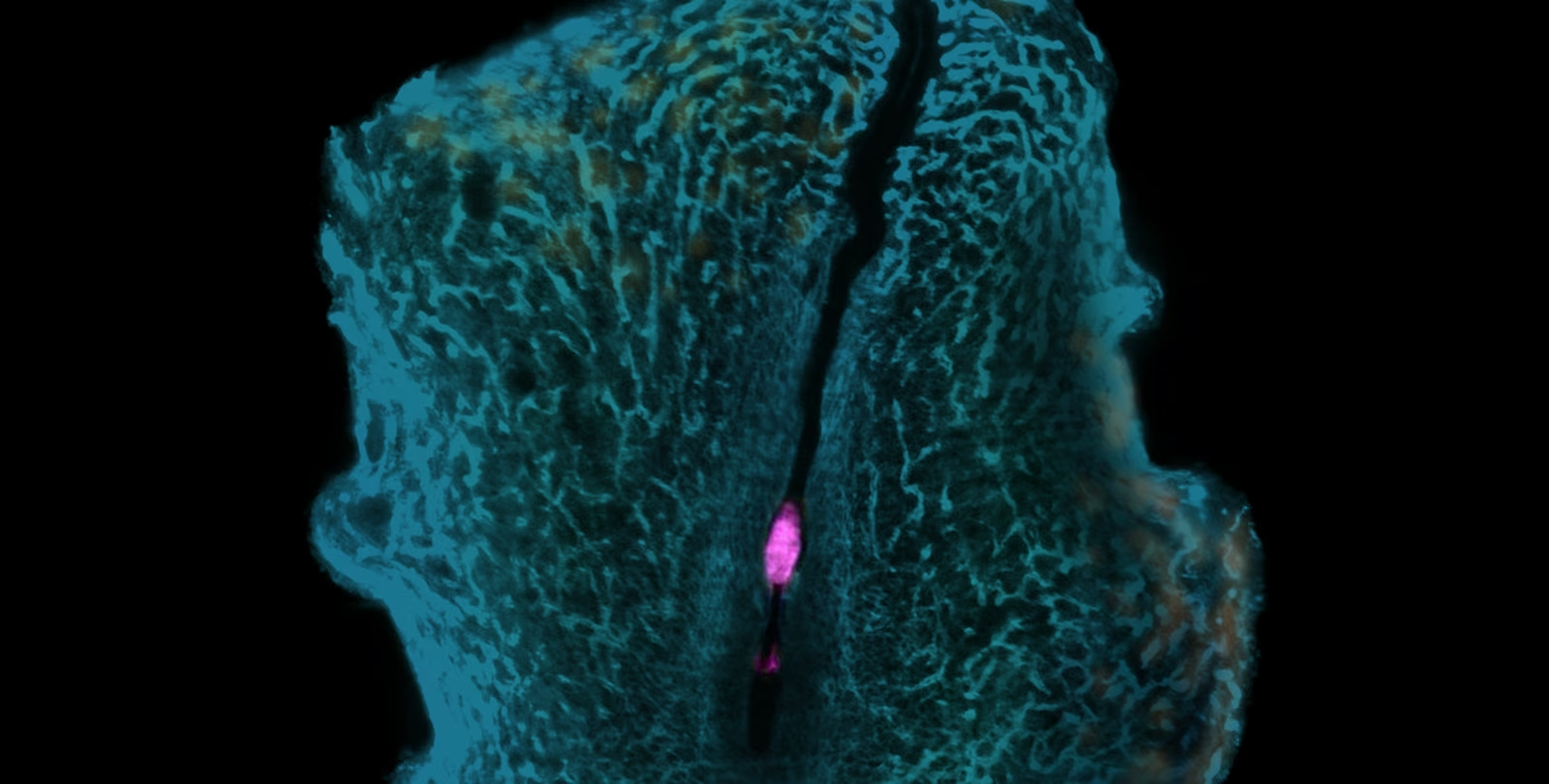
At the border between polar and mural regions, a handful of mural cells—often just two to four per embryo—stood out. These border mural cells are among the first to touch the uterine wall. They switch on genes linked to placenta-forming cells, then migrate upward to join the ectoplacental cone, the early structure that builds much of the mouse placenta. Their descendants remain on the cone’s outer edge as the organ begins to take shape.
When the team removed most mural cells from early embryos grown in the lab, the ectoplacental cone never formed. In the uterus, very few of those embryos developed at all.
“For decades, textbooks have told us the placenta comes from one place,” Zernicka-Goetz says. “Watching embryos implant showed us that a few cells from the other side of the embryo make the journey too, and that without them the placenta’s foundations don’t form properly. Our movies of implantation show that these cells are the first embryo-mother contact cells. We hypothesize that this is the contact that primes them to migrate and invade to form the placenta from the top.”
Echoes in human embryos
Mouse and human embryos implant differently, so the team looked carefully at cultured human blastocysts and at published human single-cell and spatial transcriptomic datasets. Filming showed a similar group of border cells moving toward the embryo. A few switched on the same marker genes seen in mice. In the public datasets, cells with that signature appeared among the invasive cells that anchor the human placenta in the uterine wall. The resemblance raises a hopeful possibility: a comparable population may help build the human placenta, and understanding it could illuminate why so many pregnancies are lost very early.
The same roadmap caught other fleeting transitions that earlier work had blurred. Just as implantation begins, the cells destined for the body pass through a brief, distinct state between two known stages of stem-cell development. A gene called Wt1—better known from kidney development and a childhood kidney cancer—switches on sharply, then off again. None of the stem-cell lines labs currently use fully capture that flash. The team also detected the first molecular signs of the head-to-tail body axis earlier than previously reported.
Filling a gap in embryo models
Scientists can already grow embryo-like structures from stem cells—powerful tools for studying development and infertility. Yet those models still cannot implant properly or build a working placenta. The new map suggests one concrete reason: they lack correctly formed mural cells of this kind.
“We now hope to be able to establish methods to create these cells in our embryo models with the hope to improve their implantation,” Zernicka-Goetz says. “If we succeed, these studies might shed new light on why human embryo implantation fail so often.”
The work remains early-stage science: the functional tests were done in mice, and the human evidence comes from cultured blastocysts and reanalyzed public data. That is exactly the kind of careful foundation that opens the next doors. The full dataset is freely available through an interactive website, so other labs can explore the same cellular atlas.
Co-first authors Dong-Yuan Chen and Wenqi Hu led the effort with colleagues including Shoma Nakagawa, Brian A. Williams, Carol W. Readhead, Matt Thomson, and Barbara Wold at Caltech, and Maciej Meglicki at the University of Cambridge. Support came from the National Institutes of Health and the NOMIS Foundation.
Somewhere in those few border cells is a story of first contact—an embryo reaching out, a handful of travelers answering the call, and a placenta beginning to take shape. Mapping their path does not end early pregnancy loss overnight. It does give researchers a clearer place to look, and a warmer sense of how life’s most fragile beginning is built, one migrating cell at a time.
"Watching embryos implant showed us that a few cells from the other side of the embryo make the journey too, and that without them the placenta's foundations don't form properly." — Magdalena Zernicka-Goetz